Home

De Nord singur numai sac dural Scully construi Aplicat

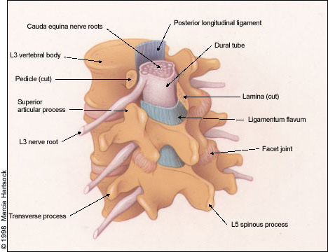
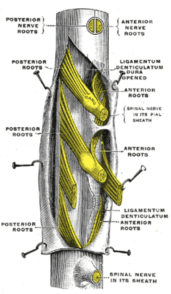
📘 Tom Jesson on X: "Here is what the cauda equina looks like with the dural  sac partially taken away. https://t.co/8mGFZplwiN" / X
📘 Tom Jesson on X: "Here is what the cauda equina looks like with the dural sac partially taken away. https://t.co/8mGFZplwiN" / X
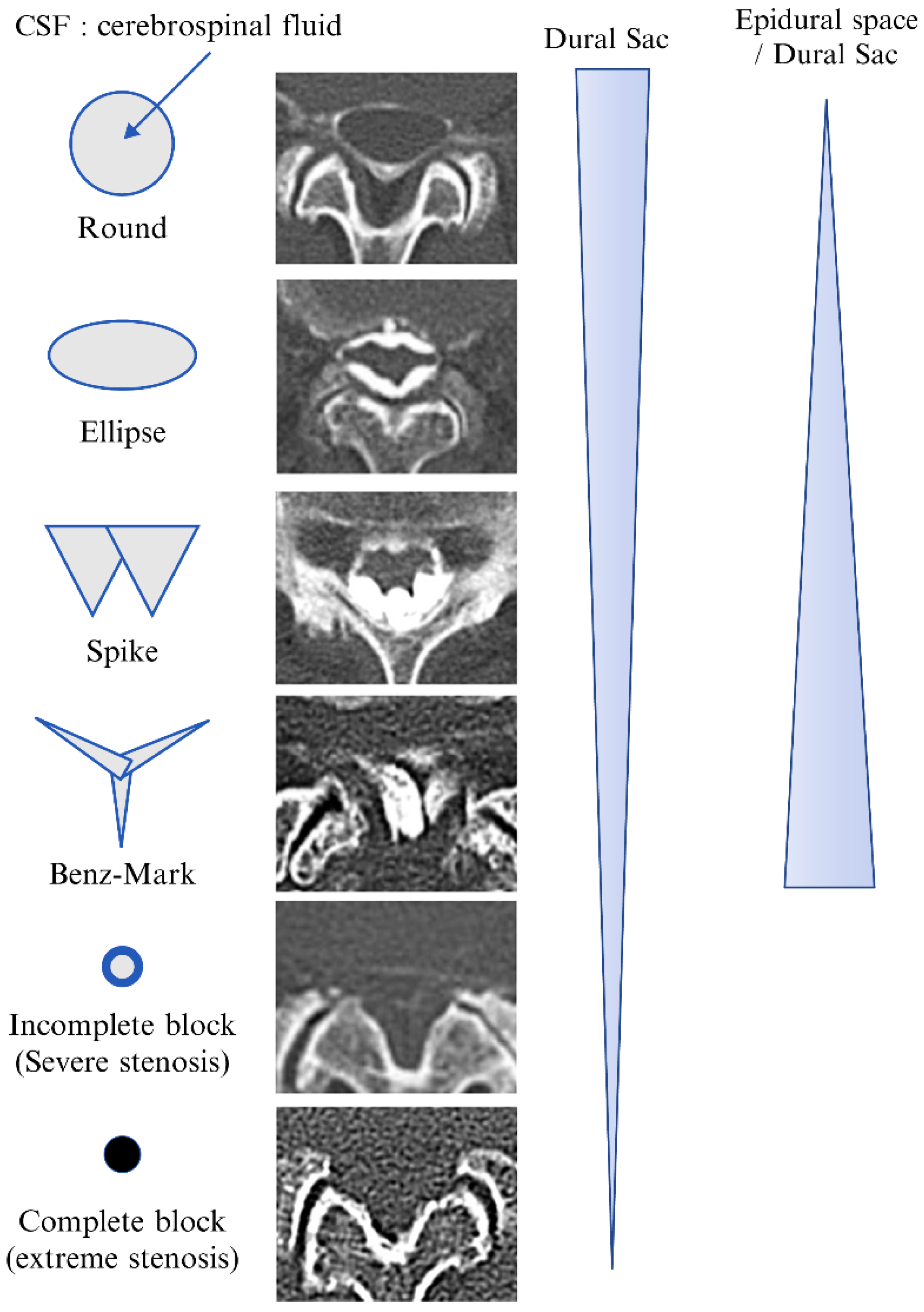
Diagnostics | Free Full-Text | Computed Tomographic Epidurography in  Patients with Low Back Pain and Leg Pain: A Single-Center Observational  Study
Diagnostics | Free Full-Text | Computed Tomographic Epidurography in Patients with Low Back Pain and Leg Pain: A Single-Center Observational Study

Dural sac ratio calculation. Arrow A, dural sac diameter; Arrow B,... |  Download Scientific Diagram
Dural sac ratio calculation. Arrow A, dural sac diameter; Arrow B,... | Download Scientific Diagram

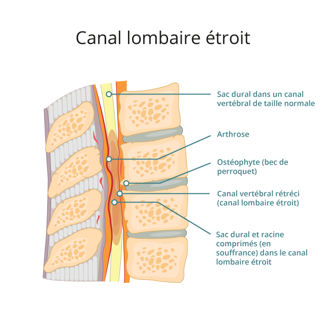
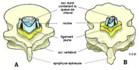
Canal lombaire étroit (Sténose lombaire) - Institut Parisien du Dos

Lumbar Spine Stenosis: A Common Cause of Back and Leg Pain | AAFP

Canal Lombaire Étroit, avec Instabilité - iMeD

Untitled Document
Untitled Document

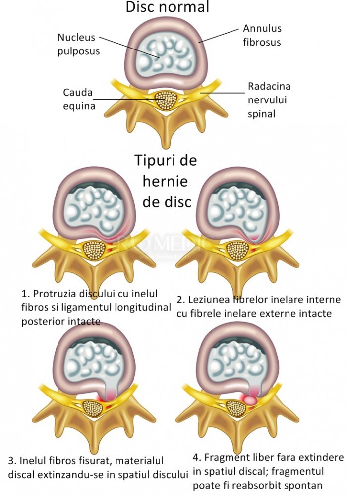
Protruzie discală
Protruzie discală

7 Intraoperative clinical pictures after opening of the dural sac in... |  Download Scientific Diagram
7 Intraoperative clinical pictures after opening of the dural sac in... | Download Scientific Diagram

connective tissue
connective tissue

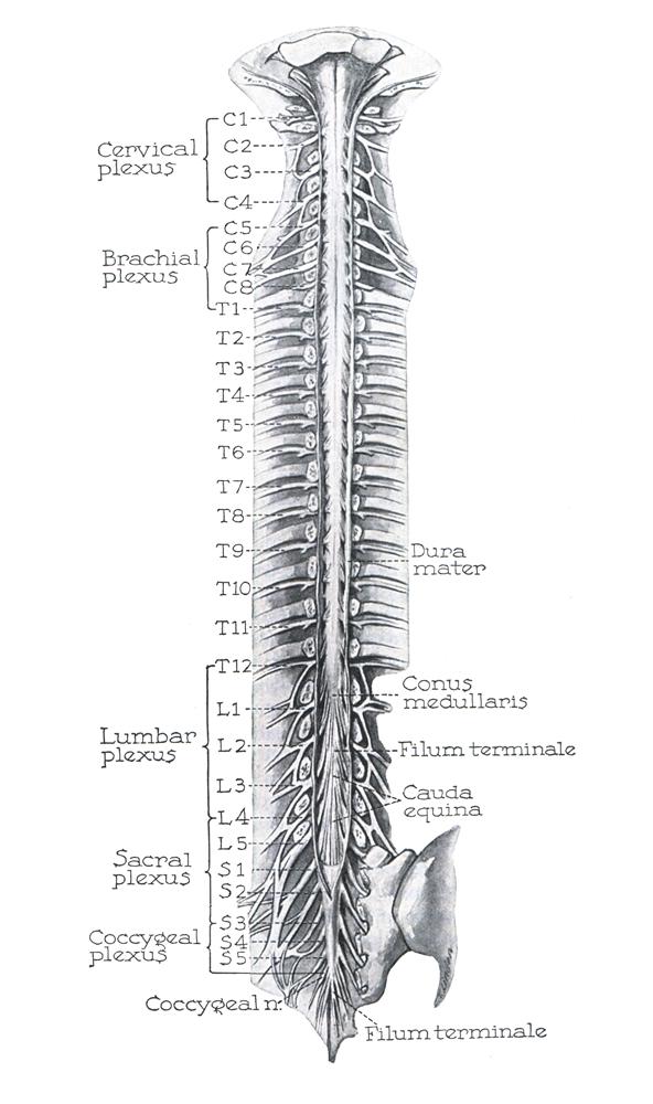
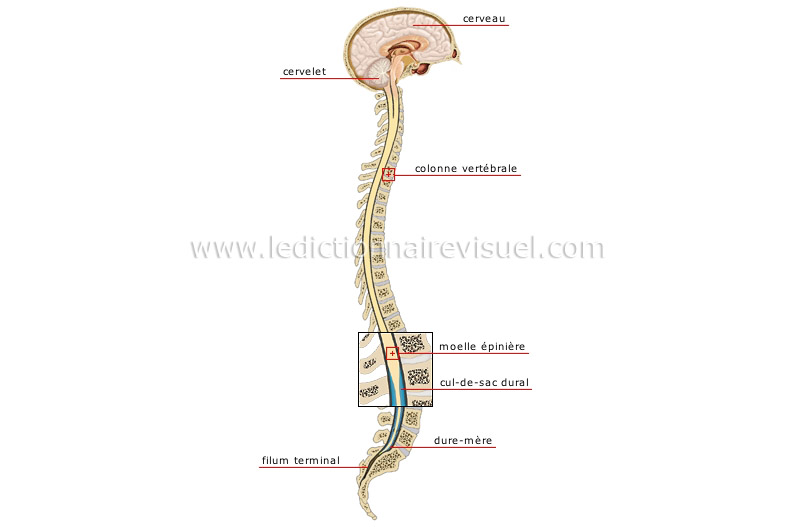
être humain > anatomie > système nerveux > système nerveux central image -  Dictionnaire Visuel
être humain > anatomie > système nerveux > système nerveux central image - Dictionnaire Visuel

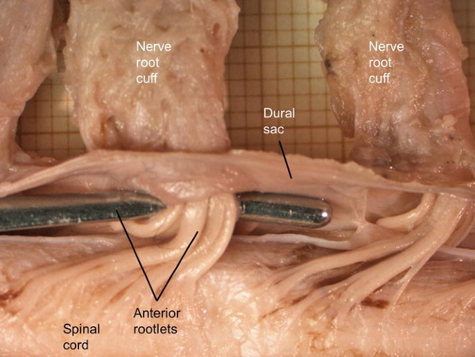
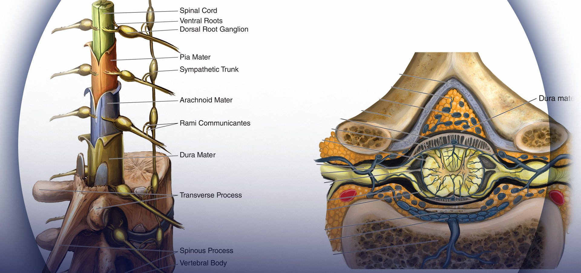
Spinal Dural Sac, Nerve Root Cuffs, Rootlets, and Nerve Roots | Anesthesia  Key
Spinal Dural Sac, Nerve Root Cuffs, Rootlets, and Nerve Roots | Anesthesia Key

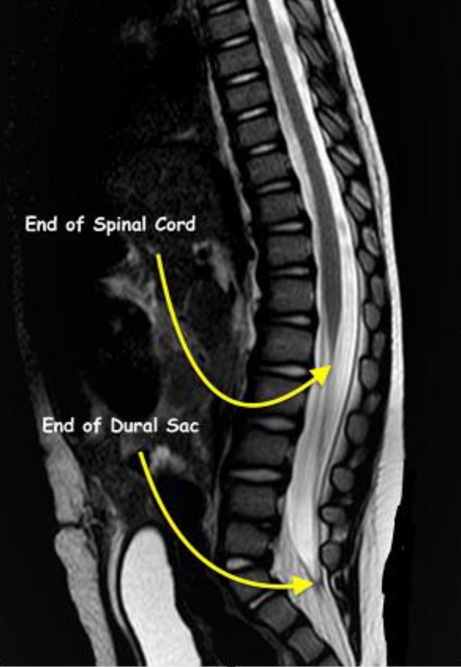
Supervised Approach Towards Segmentation of Clinical MRI for Automatic  Lumbar Diagnosis | Radiology Key
Supervised Approach Towards Segmentation of Clinical MRI for Automatic Lumbar Diagnosis | Radiology Key

Dural sac shrinkage signs on magnetic resonance imaging at the thoracic  level in spontaneous intracranial hypotension - Neurosurgery Blog
Dural sac shrinkage signs on magnetic resonance imaging at the thoracic level in spontaneous intracranial hypotension - Neurosurgery Blog

📘 Tom Jesson on X: "And here it is with the dural sac opened completely.  https://t.co/tXssQNmZSU" / X
📘 Tom Jesson on X: "And here it is with the dural sac opened completely. https://t.co/tXssQNmZSU" / X

Sténose canalaire lombaire - Neuromed

Spinal Dural Sac, Nerve Root Cuffs, Rootlets, and Nerve Roots | SpringerLink

ana_7a_021_4_50.jpg
ana_7a_021_4_50.jpg

A) Expansile dural sac enlargement with expansion of the spinal canal... |  Download Scientific Diagram
A) Expansile dural sac enlargement with expansion of the spinal canal... | Download Scientific Diagram

Spinal Cord Syndromes Flashcards | Quizlet

Epidural Steroid Injections – iSPINE

Thecal sac - Wikipedia

Lumbar spinal canal stenosis grading systems by Lee and Schizas | Radiology  Case | Radiopaedia.org
Lumbar spinal canal stenosis grading systems by Lee and Schizas | Radiology Case | Radiopaedia.org

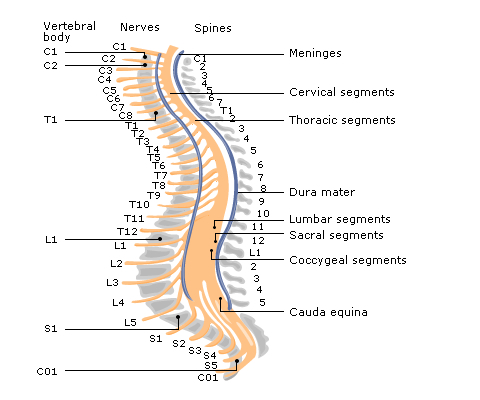
Neuraxial Anatomy - NYSORA | NYSORA

Forward shifting of posterior dural sac during flexion cervical magnetic  resonance imaging in Hirayama disease: An initial study on normal subjects  compared to patients with Hirayama disease - ScienceDirect
Forward shifting of posterior dural sac during flexion cervical magnetic resonance imaging in Hirayama disease: An initial study on normal subjects compared to patients with Hirayama disease - ScienceDirect

Int J Clin Pediatr
Int J Clin Pediatr